|
After a recent cold front moved in and
dumped about two inches of snow in the Payson area, I went out
to the East Fork of the Verde River to the north to see if any
microscopic life was left in the river during the cold 25 degree
weekend day. The river was mostly frozen over and nothing but
a brown dead bottom was seen through the transparent ice. But
then I found a small area of green under the ice! A small algae
patch was still growing in one small area, about 3 feet in size.
I broke a small hole in the ice and reached in with the forceps
and pulled out a teaspoon sized sample. What would it contain?
Would anything be moving? Much to my surprise, the algae patch
was full of microcsopic life wintering the cold in their own
oasis. Here are the images I took when I got home and defrosted.
Click
on all small images for the larger 1290 x 960 sized image.
60x Images
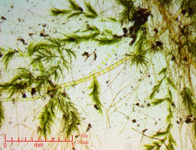 |
The algae
was a tightly interlocked and had a fern like appearance. We
saw this last month in greater profusion when it was much warmer.
Mixed in were a few isolated strands of solitary algae, and at
this magnification, little else was seen. Compare this rather
bland view (transmitted light) with the dark field illumination
of the same field below...
(Field of view
here about 3mm wide)
|
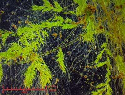 |
The dark field
was amazing! Not only did this type of illumination bring out
the rich colors of the algae, but the dark background revealed
hordes of super small organisms swimming all arround. It was
full of tiny life.
You can see why
I like shooting dark field, its so awesome for the lower magnifications.
|
150x
Images
 |
The
dark field can also be used with the 10x objective as well (Yeilding
150x), and shows intricate details in the algal cells. Most of
the tiny blips are non moving bacteria or loose debris. Moving
protists are not seen in focal stacked image sets like this. |
 |
Like
looking at the ferns in a forest. Except these are 1/10 of a
millimeter long. You can just start to see individual cells in
the ferns. |
|
Movie
Clip 1 |
A
sized down Youtube clip I posted at this 150x magnification.
The swarm of swimming protists are like fire flies at night!
The original movie was 1790 wide (half sized), and would have
taken 2 hours to upload... |
600x
Images
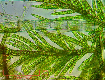 |
We change
to brightfield back lighting for higher magnifications. (darkfield
doesnt work with the higher power objectives I have)
Here the algal
strands show rich internal details. A small greenish protozoan
can be seen on the extreme upper left corner.
|
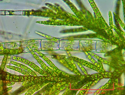 |
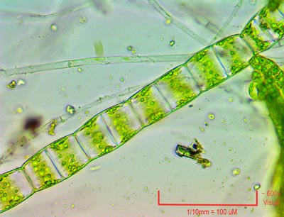
Centering
the solitary algal strand. The small green chloroplasts can be
seen within. The partitions between cells is very evident. |
 |
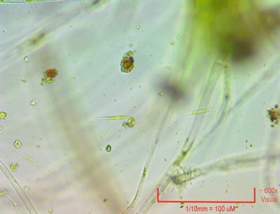
Another
type of algal strand showing the clear cell with green chloroplasts
internally. Small protozoans flit in and out of focus here, despite
being under a microscope cover slip. |
 |
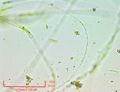
Those
small greenish rod shapes that were slowly traveling from left
to right are freshwater diatoms. They are protozoans filled with
green chloroplasts to convert sunlight to food. They are far
more rare this time of year in the river. They have skeletons
composed of pure silica quartz. |
 |
A
few stationary protists and algal strands. |
 |
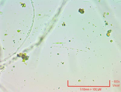
In
the center is another type of diatom. As you can see, diatoms
are very small. Look to the lower right and there is another
spear shaped diatom. |
 |
Centering
that diatom. The small round spheres of protoplasm are bacteria. |
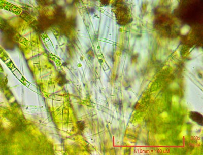 |
Colony
of algae and protists having a party. |
1500x
Images (Oil Immersion Objective)
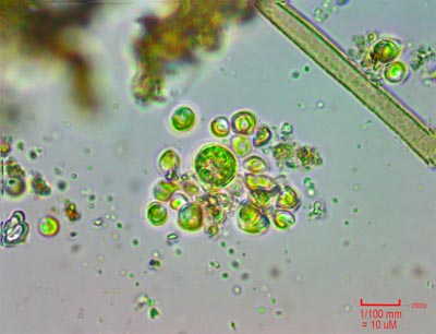 |
Challenging
indeed to image slowly moving protists at this magnification!
But if they stand still long enough, you can get splendid internal
details at this power. Here is a small cluster of cells. |
 |
This
protist wouldnt stop moving, but I tried. |
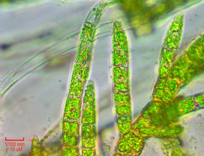 |
Fern
like algal fronds tips showing internal cell details. |
 |
Two
types of algal filaments. Arcing across the top is a smaller
type with amazing internal details in the cells. The one big
single cell of a strand is in the center. Its filled with a yellow
type of chloroplast. Chloroplasts come in two flavors - green
and yellow. |
|
Camera:  10 Megapixel CMOS
Platform:  AmScope Trinocular 2000x
Filters:  NONE
Location:  Payson, Az Verde River
Elevation:  5200 ft.
Processing: Photoshop  CS Pro
HOME


