|
A variation on dark field illumination
adds the element of color contrast to transparent objects that
are difficult to image clearly any other way. While dark field
gives you a black background and glowing subject, you can increase
the visibility even more on difficult to image subjects such
as totally clear protozoans or very clear crystals. The idea
behind Rheinberg illumination is to give the background and subject
contrasting colors, such as a blue background and yellow back
illumination. Here are some experiments with granular sugar crystals,
which I found to be not only cubic but some are rectangular in
shape as well, unlike salt which is always cubes.
Please click on
images for the 1290 x 960 images. (Reduced from the 10 megapixel originals)
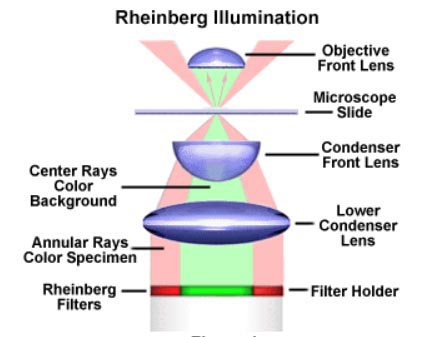 |
Graphic
from the web site Molecular Expressions showing how Rheinberg
illumination works. A bi colored filter is inserted in the condenser
and projects two cones of colored light upward. The inner cone
will be the background, and the backlighting for dispersion is
the ring of light outside the inner cone. Ill show you some actual
microscope stage shots to show you how this actually works. |
 |
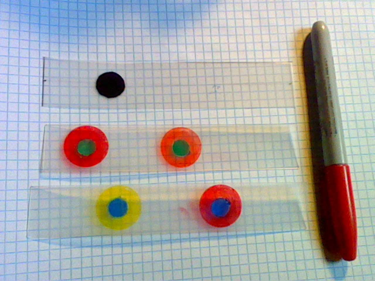
Rheinberg
masks are made by using colored markers or cellophane on a sliding
plastic base to go into the side of the condensers. They provide
a slit for this for both dark field illumination ( upper piece)
and several types of Rheinberg illumination. The central disk
is the background color, and the ring outside of it will become
the back illumination for the transparent subject. |
 |
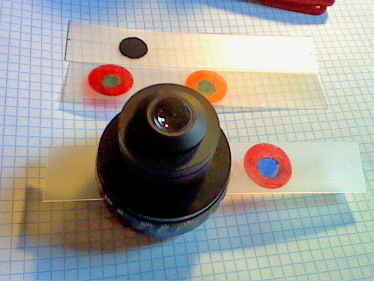
Slid
into the condenser and ready to go. |
 |
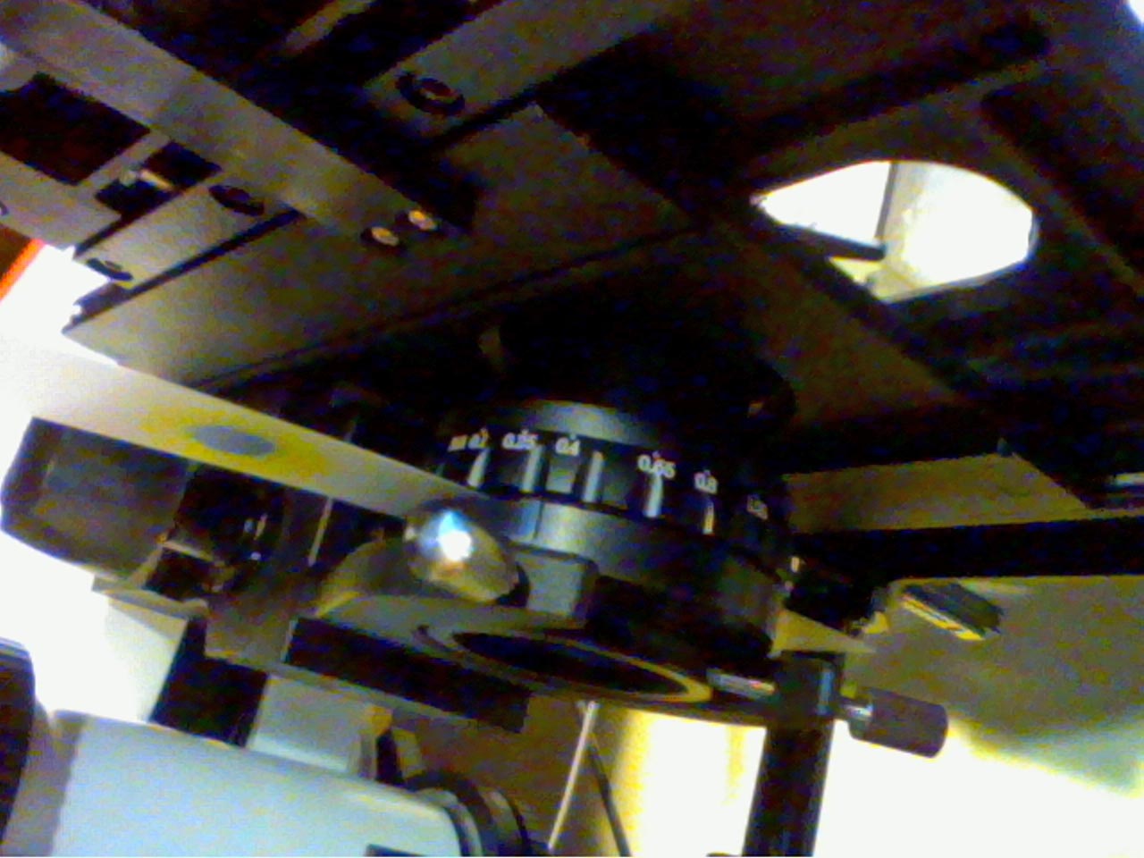
Under
the microscope stage we mount the condenser, and you can see
the plastic strip with the colored areas inserted. |
 |
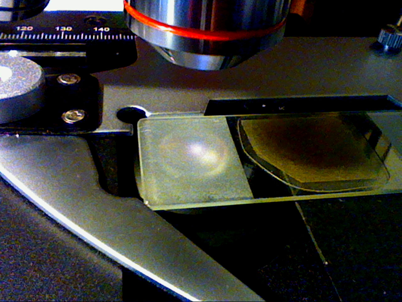
It
is quite hard to get a clear shot of what Rheinberg illumination
looks like. Here, the condenser is in focus and concentrates
the outer ring to a sharp pin point on the specimen. But you
cant see the inner blue cone until you do this.... |
 |
By
lifting the slide (which is frosted for clarity) up, the blue
cone becomes visible. THIS is what the microscopes objective
lens sees - a blue background but not the yellow ring. |
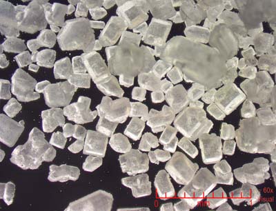 |
This
is a standard dark field shot (using only a black mask for the
central disk and a clear ring around to back illuminate) Sugar
at 60x shows a variety of shapes, all cubic and many are rectangular. |
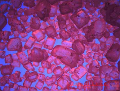 |
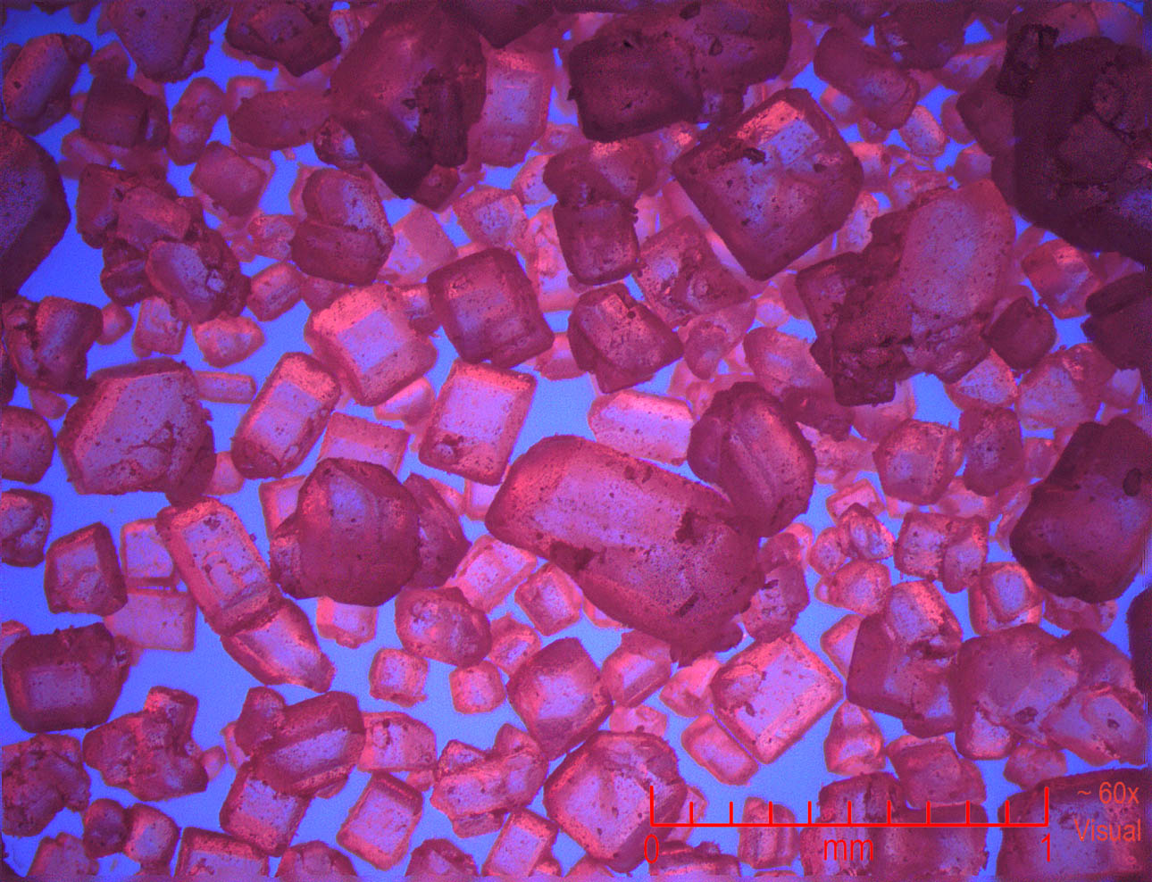
With
Rheinberg Illumination, we here use a blue center surrounded
by a red ring. Note the nice color contrast of the crystals now!
|
 |
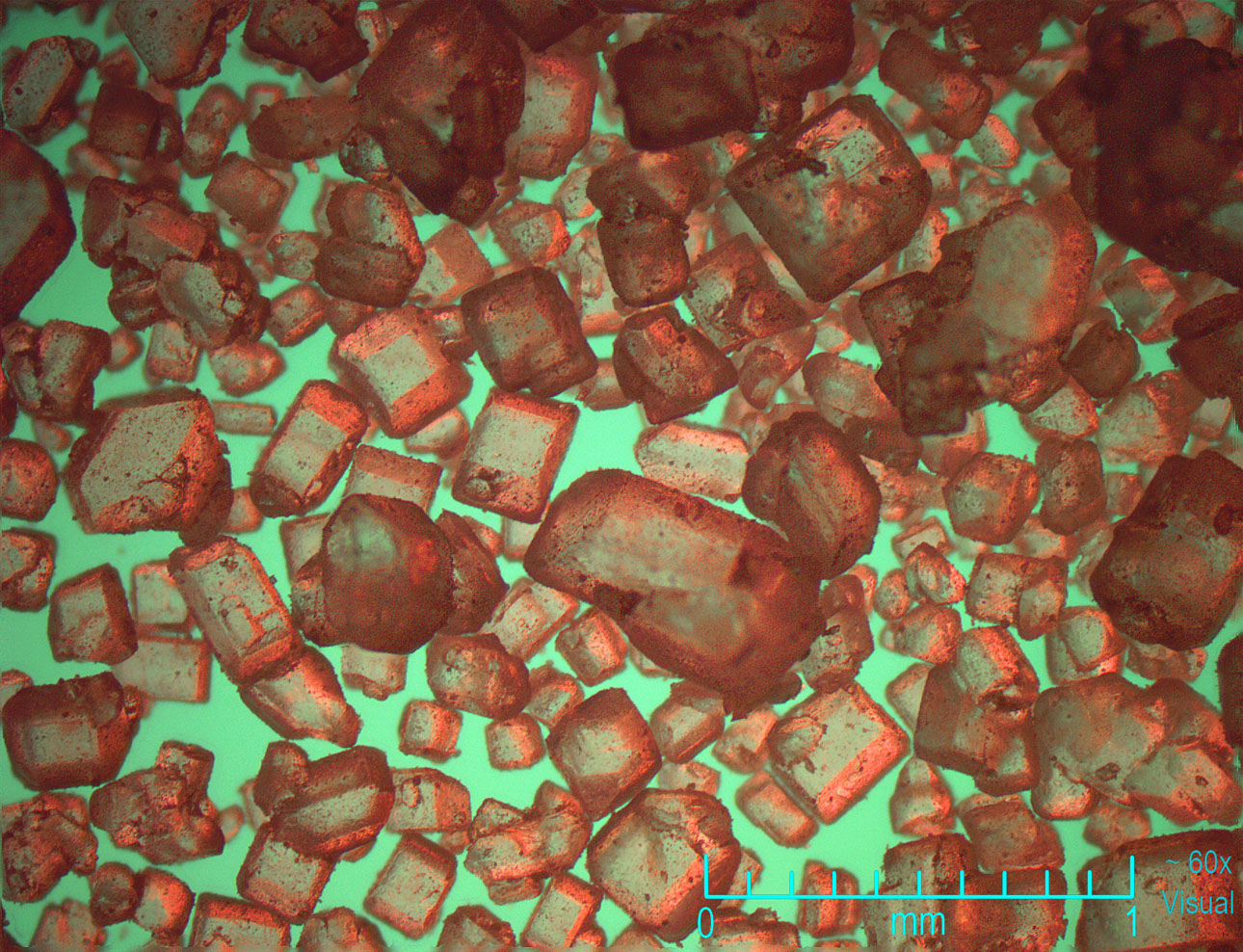
Using
a green disk and red ring did not work quite as well but is interesting.
60x |
 |
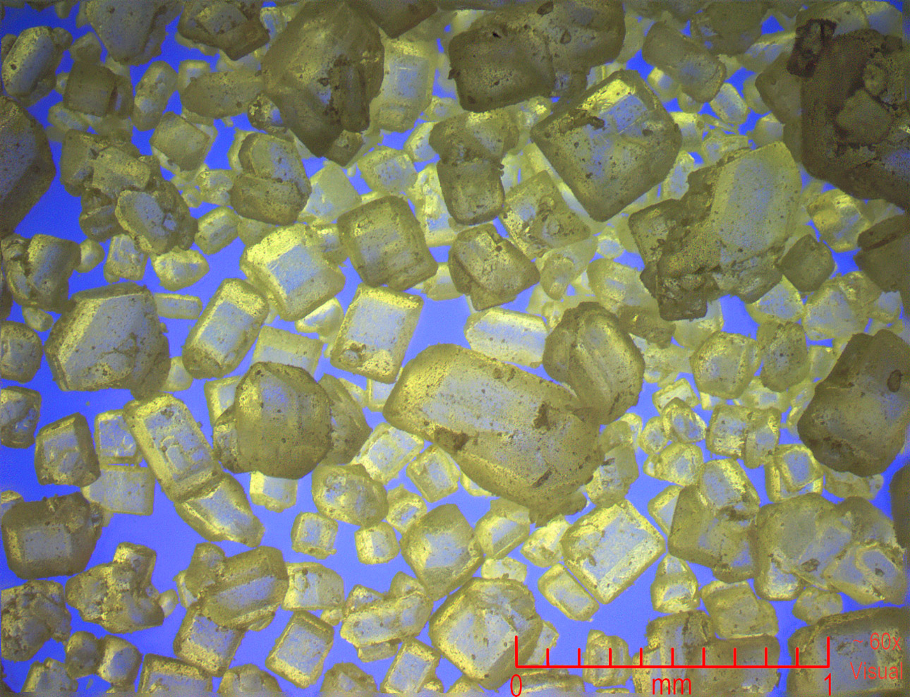
Better
is the most used colors - blue background (center disk) and yellow
back illumination (outer ring). 60x
Small clear marine
and freshwater invertebrates show up very well with this color
set, and look natural too.
|
 |
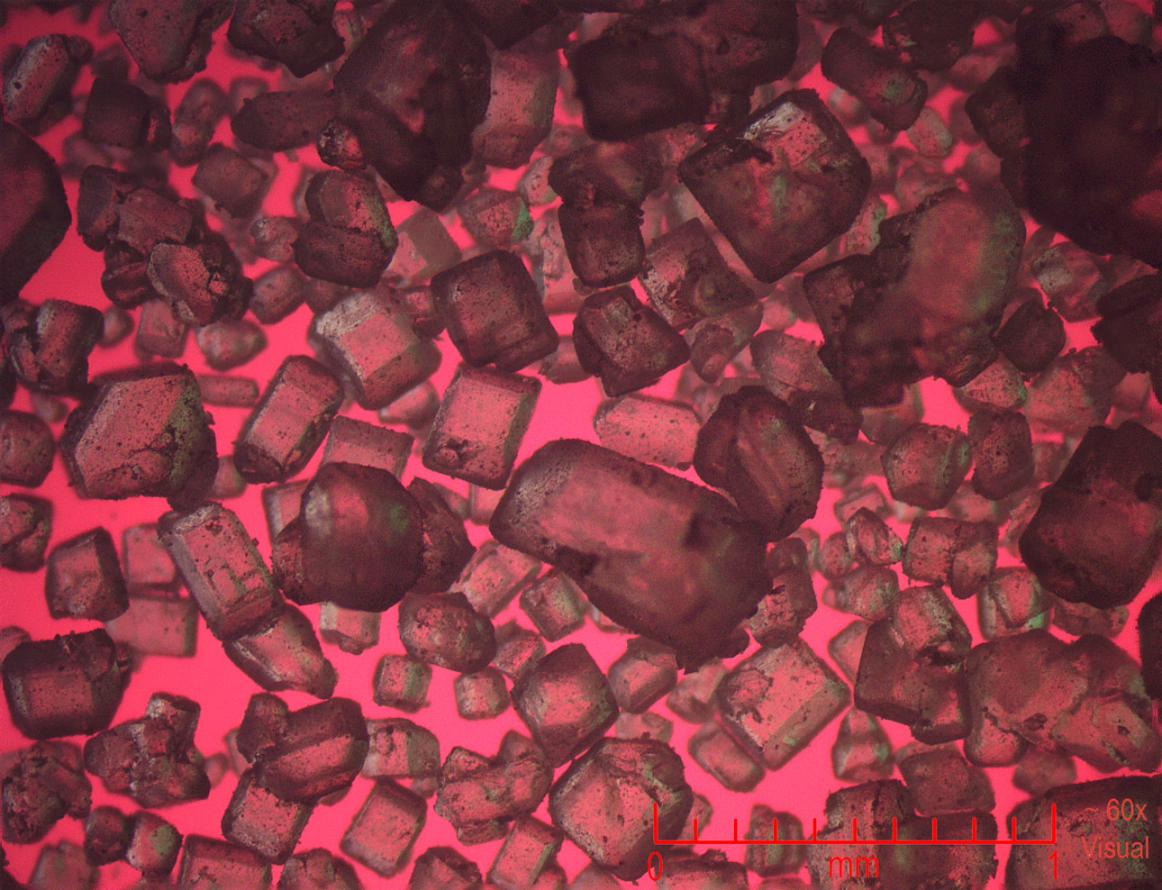
The
red background and green ring was not as nice. |
 |
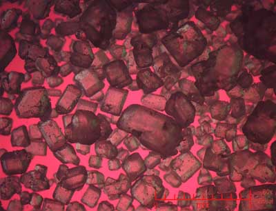
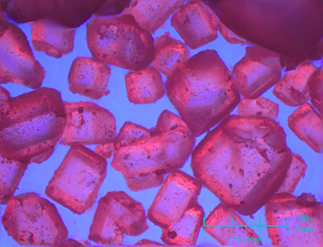
Now
we move up to the 10x objective yielding around 150x on this
image. This of course is a dark field shot of the crystals. |
 |
A
blue background and red ring makes the shot far more exciting
and more detail can be seen. |
 |
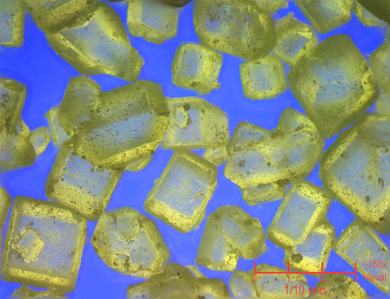
A blue
background and yellow back illumination is very clear as well.
In summary, Rheinberg
illumination will be come a powerful tool in the future for my
imaging work and I am looking forward to trying it out on many
other materials and living things!
|
|


Camera:  10 Megapixel CMOS
Platform:  AmScope Trinocular 60-150x
Filters:  Rheinberg
Location:  Payson
Processing: Photoshop  CS Pro
HOME