|
During my 8 mile run this morning, I passed
the School yard track and spotted a low area with standing stagnant
water from all the rain we had in October. Since I carry a specimen
vial with me in my running pack (dont we all?) I brought back
a teaspoon of water to examine under the microscope back home.
As bad as the water looked, there was little lively activity
in it. Only a few swimming protozoans, a few isolated algae strands,
and some clusters of some sort of cyanobacteria. Closer examination
of this apparently meager specimen showed some very interesting
things to see!
Please
click the thumbnails below for the full size view.
600x
views:
 |
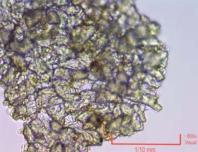
These
clumps of transparent cells appear to be some form of cyanobacteria.
The bottom of the basin was covered with this material which
appeared brownish to the eye. Here at 600x the separate cells
can be seen. Mixed in were spores, some round ball like algae,
a few swimming protozoans, and some other very strange things. |
 |
Seen
here is one of the algae spheres. It is in the very center and
stuck to the cellular mats. I could see structure inside, so
a closer view was warranted! |
 |
A
solitary strand of algae. You can see some remarkable things
here. The cell walls partitioning the strand are clear, and inside
each cell some amazing things were going on. A series of green
patches in each cell for the photosynthesis part, and moving
dark colored spheres were bouncing around inside as I watched.
I had never seen anything like this before. |
1500x
views:
 |
Now
lets jump to the 100x oil immersion objective for the remaining
shots for a combined magnification of 1600x. The close ups revealed
so much more details! This is the solitary algae strand. The
green patches are now clear, and look inside each cell. See the
small black spheres? They were moving around rapidly inside.
I think this was due to Brownian motion rather than swimming
but it looked amazing. |
|
Movie
1 |
Here
is a You tube movie I made for 16 seconds of the action in the
above shot. Watch what is going on inside an algae cell. |
 |
One
of the round algae spheres. The internal detail was unexpected.
Here at 1600x you can see the organells inside and the sharp
cell wall. On the left is some sort of bacterium and to the upper
right more bacteria in a cluster. |
 |
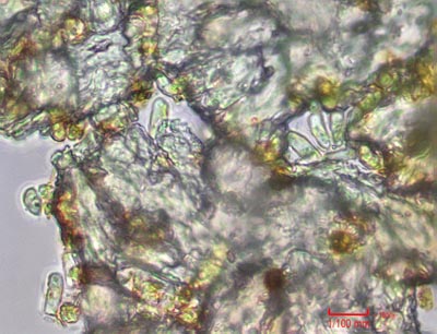
Another
area of the cyanobacteria mat, showing several bacteria mixed
in. |
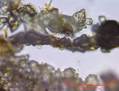
Now this one
was very peculiar! This apparent algae strand was SWIMMING along
in a sinuous line between pieces of mat. Individual cells are
linked in this worm like algae. I have no idea what it is.
|
Camera:  10 Megapixel CMOS
Platform:  AmScope Trinocular 40 -2000x
Filters:  NONE
Location: Payson, Az
Elevation:  5100 ft.
Processing: Photoshop, Picolay
HOME


