|
Today, we took another trip to the Payson
Recreational Lake, and now that the huge algae bloom is over
with, I sampled areas around the base of cattail water plants
and areas near the dock. A huge change in fauna and plant life!
I also found a duck feather and found it to be most fascinating
in close up as well. Here are todays finds!
Please click each
thumbnail below for the full size 1290x960 image!
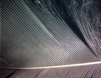 |
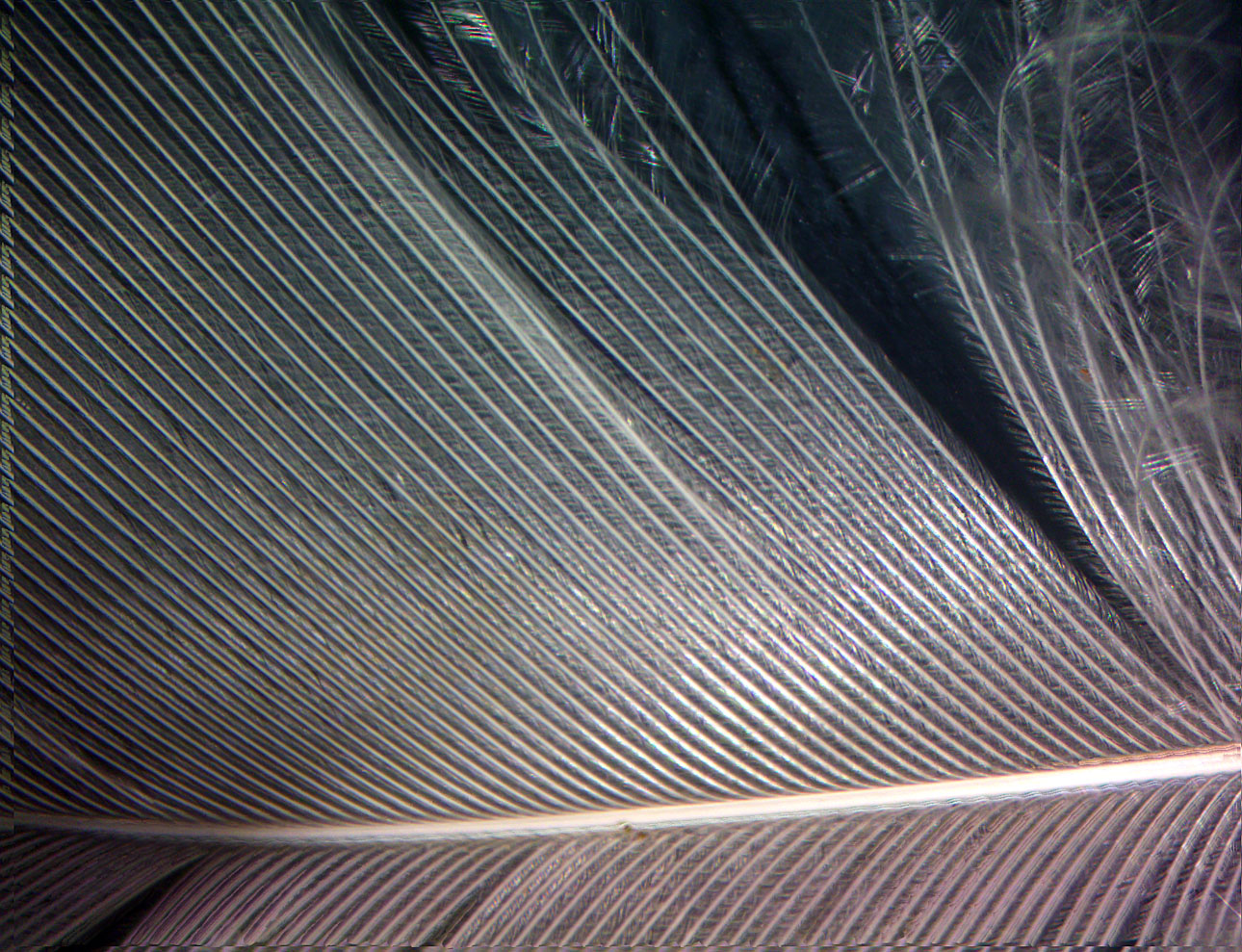
Lets
start with the binocular stereo microscope and look at that feather
at 7x. You can see separate vanes in the feather are somehow
locked together to form one flat sheet. |
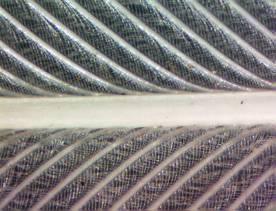 |
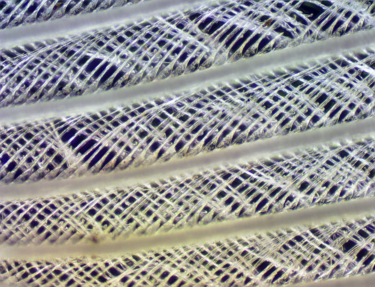
At
45x, the stereo microscope is at maximum power, and starts to
get a bit fuzzy. However you can now see the weave pattern connecting
the vanes. Now lets switch to the high powered microscope... |
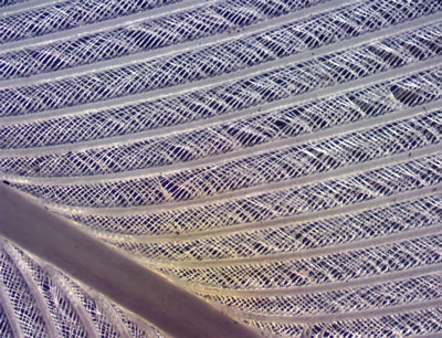 |
Using
dark field illumination on a piece of the feather, we see here
at 60x how much sharper this microscope is than the stereo. The
weave pattern is now very apparent! Remember that dark field
illumination is essentially a back lit image with a black background
achieved optically. |
 |
Our
translucent feather at 150x now shows very fine detail in the
feather vane weave. Look closely at them and you'll see hooks
and barbs starting to show up that stitches them together. |
 |
At
600x, the dark field method is at its limits, but shows now the
hooks that connect the feather vanes together. They are very
small! |
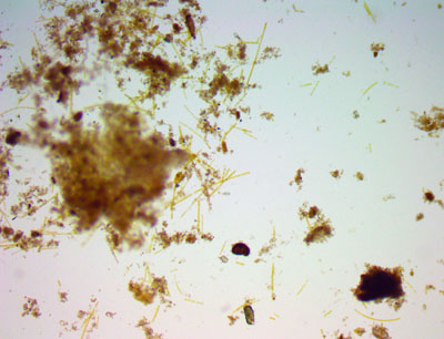 |
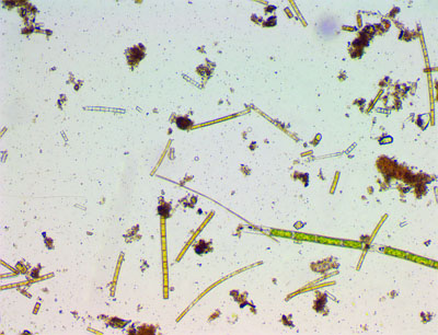
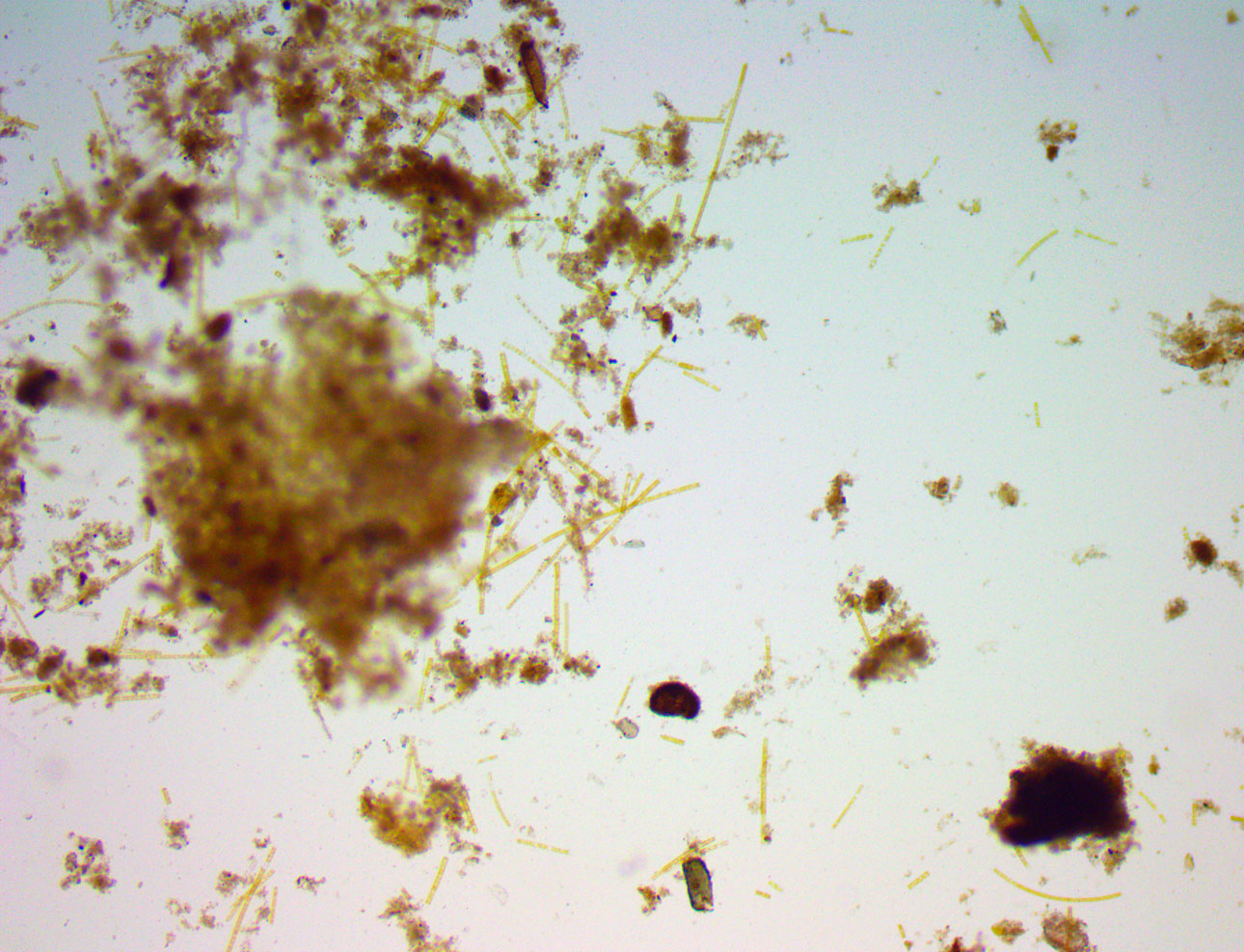
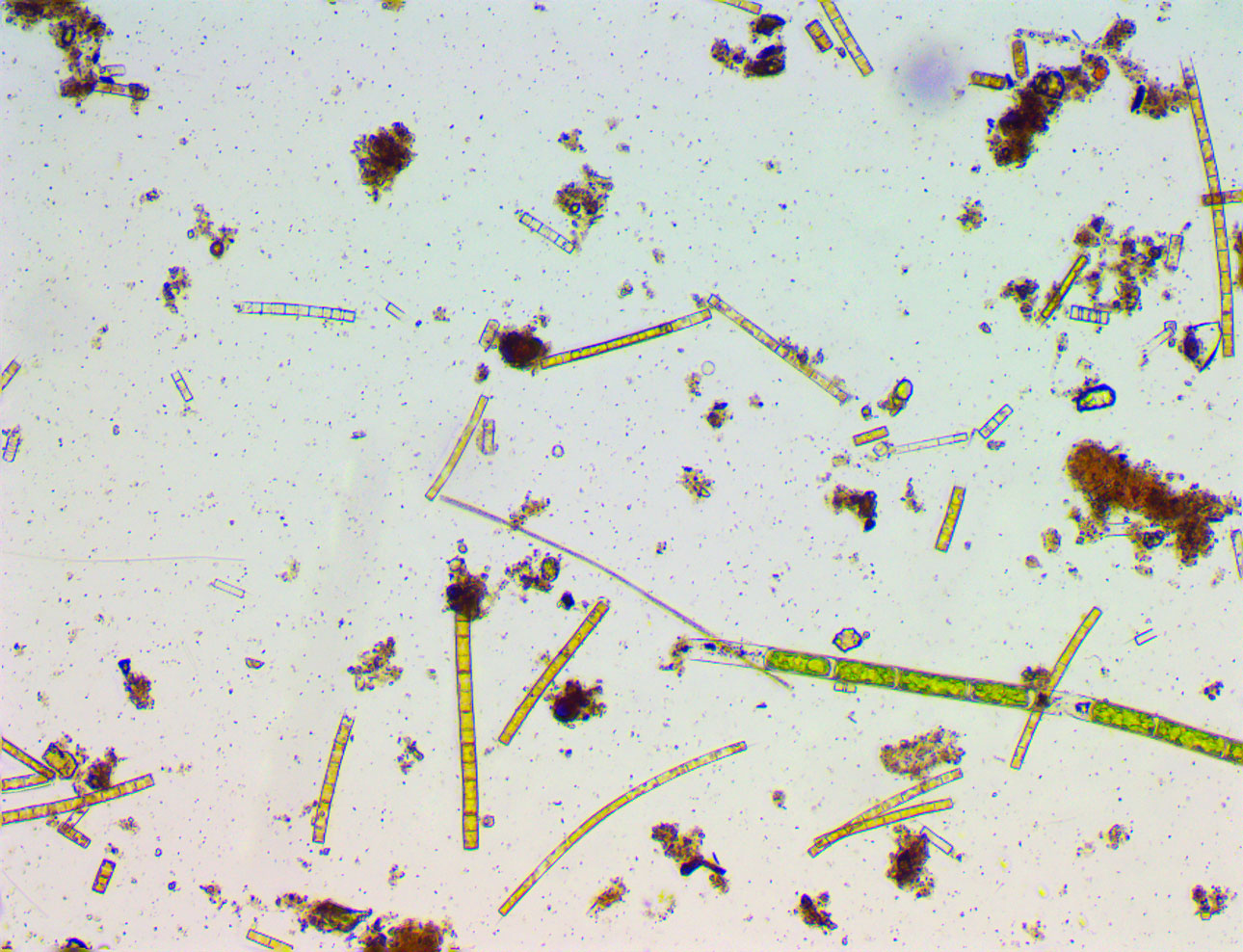
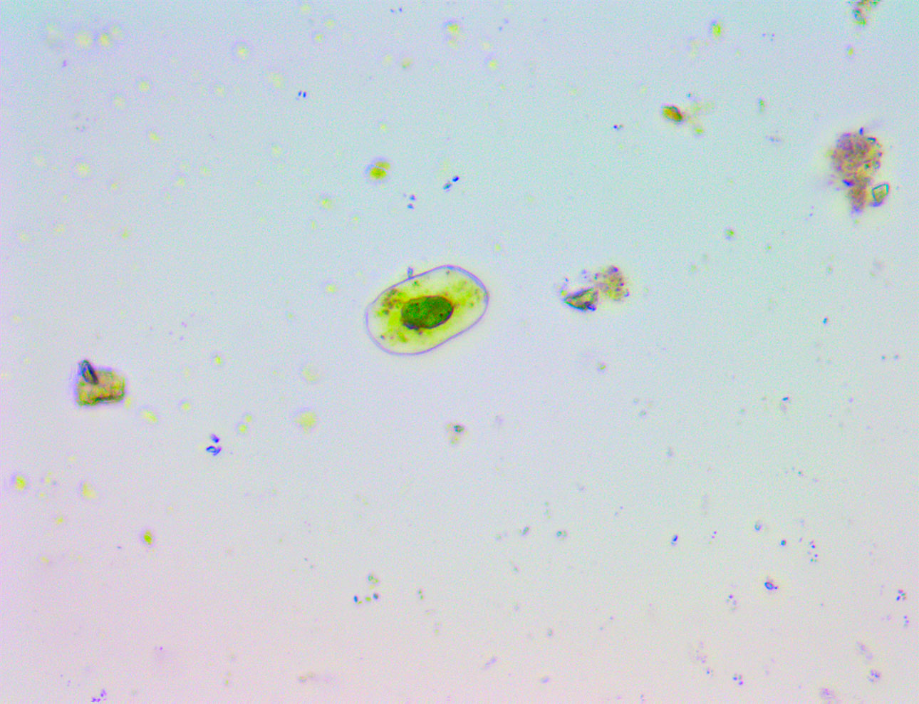
Now
lets look at that muck around the base of the cattails. At 60x,
we can see some short strands of some type of algae and organic
debris, and a few isolated round objects that have green in them.
This is a transmission light image - it was back lit like most
microscopes are set up to do. |
 |
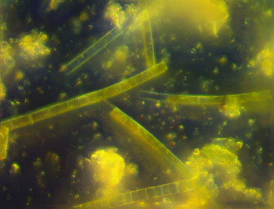
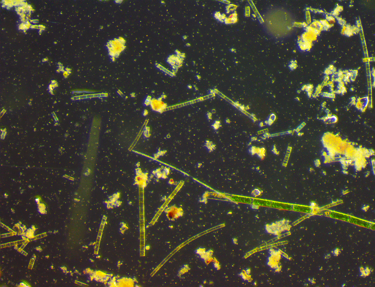
Compare
this dark field image also at 60x with the same field above.
You can see why that for low powers, the dark field method is
very exciting and shows a more natural appearance. |
 |
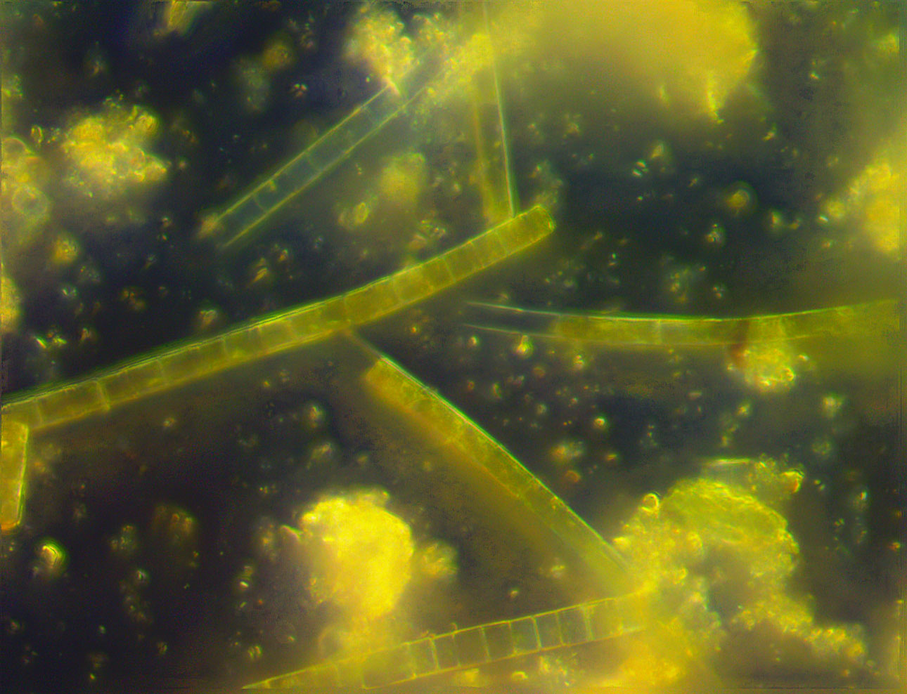
Now
lets compare two more fields, now at 150x. This close up using
transmitted light shows the algae strands as numerous short lengths.
The individual cell partitions in the strands are apparent. There
were several swimming protozoans in this shot, but stacking a
dozen frames to make the image fully sharp tends to erase them. |
 |
Dark
field view of the same 150x field. Its amazing how much more
small stuff is visible. |
 |
600x
dark field view of some of the algae strands. The dark field
technique starts to fall apart at this magnification. Its best
to use bright field. |
 |
Compare
this dark field view at 600x of an isolated algae cell with the
next image. You can see that bright field works better for sharpness
because the sub stage diaphragm can be stopped down to make the
rays more parallel. |
 |
Same
cell at 600x - but bright field transmitted light. The details
inside the cell are fantastic! |
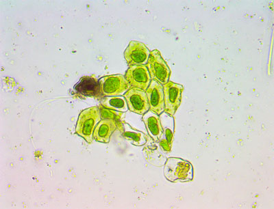 |
I
found this stunning cluster of algae cells at 600x that show
amazing details. |
 |
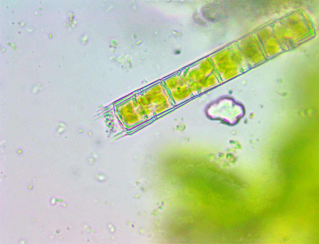
At
1500x, this algae strand shows intricate internal details. Transmission
is good for this. (Oil Immersion lens) |
 |
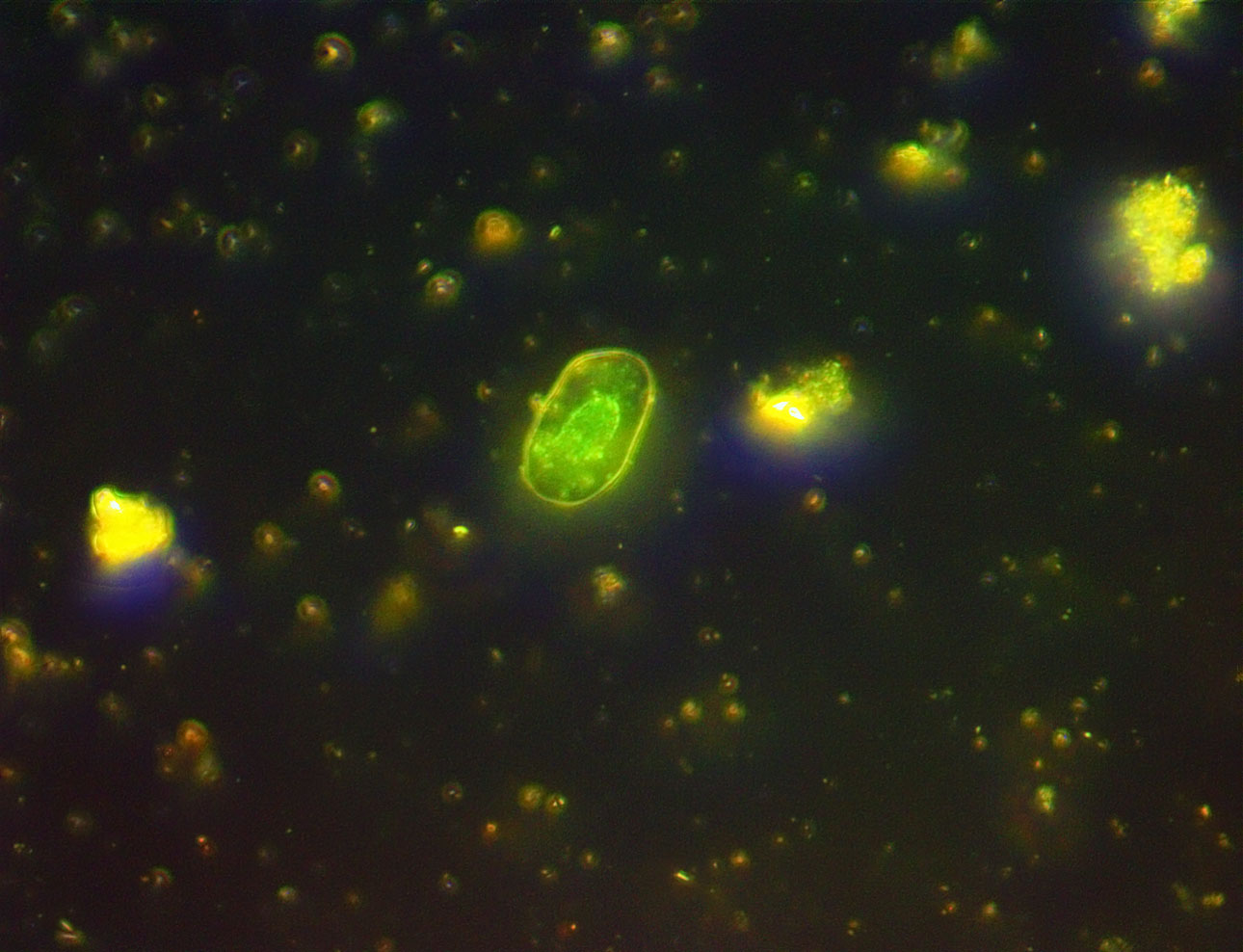
A
single frame at 1500x to try to capture one of the rapidly swimming
protozoans. This one near the top is greenish and may be a Eugleanoid.
It is very hard to capture moving animals as they dart so fast
that they move out of the tiny field very fast. The smaller round
objects here are bacteria. They dont move much, just jiggle constantly
due to Brownian motion. (Oil Immersion lens) |
 |
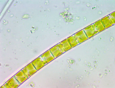
I
saw this nice algae strand at 1500x with superb internal details.
(Oil Immersion lens) |
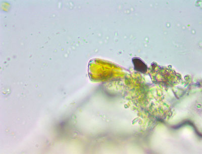 |
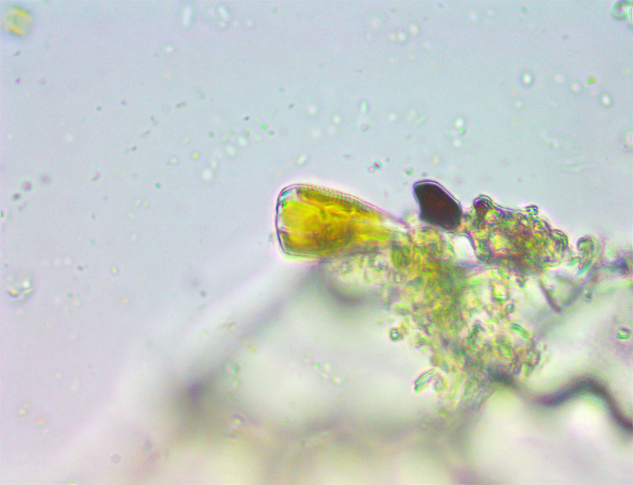
I
have no idea what this very tiny protozoan is. It is vase shaped
and at this magnification - 1500x - it shows some ribbing and
an internal wall. (Oil Immersion lens) |
|


Camera:  10 Megapixel CMOS
Platform:  AmScope Trinocular 2000x
Filters:  Dark Field stop
Location: Payson, Az
Elevation:  5150 ft.
Processing: Photoshop  CS Pro
HOME