 Microscope Images
The microcosm of the world around us

More Payson Lake Microcosm

Updated:  7/26/20


|
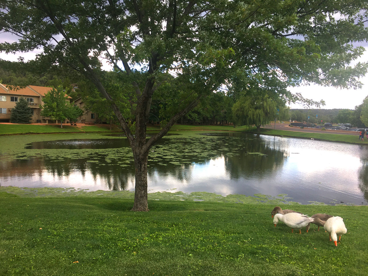
Once again on this fine summer day we traveled
to the lake in Payson to see what we could find in the algal
bloom which is currently in effect, and comes every summer about
this time. You can see in the photo above, the surface of the
lake is covered with floating algae, and duck weed. Here are
some close ups of what was found under the microscope, with a
few new surprises! All animals retuned to the aqueous environment
after the shooting session.
 |
Algal
strands, 60x. The main clots of algae formed into net like woven
arrangements. |
 |
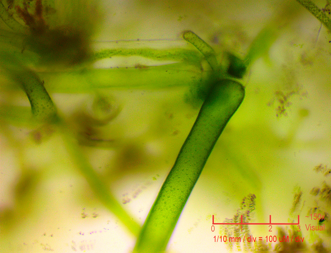
Larger
strands at 60x. Some interesting tubular algae was mixed in,
along with lots of micro life swimming about. |
 |
At
150x, we can start to see some peculiar bumps on the surface
of the larger strands. |
 |
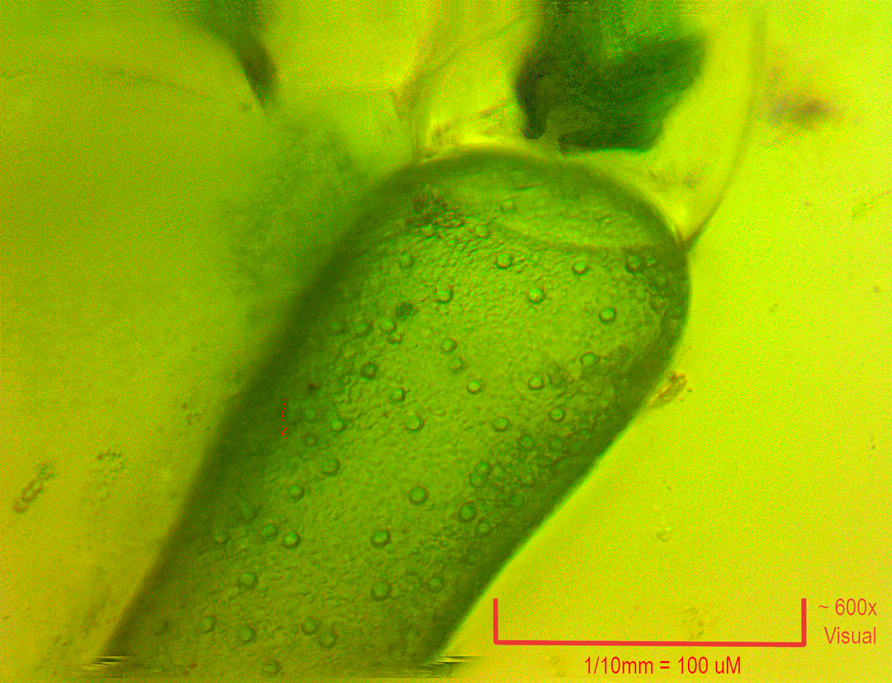
Close
up at 600x of the algal filament, showing fine details in the
bumps on the surface. |
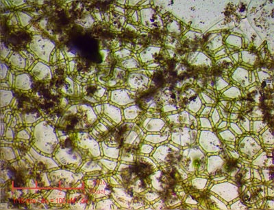 |
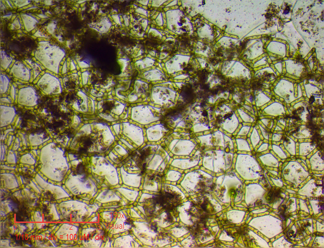
The
netlike algae at 150x. Individual cells are seen clearly. |
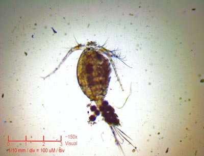 |
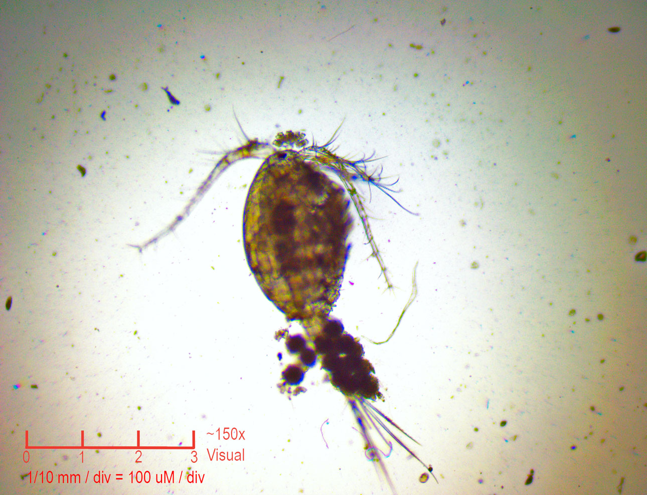
Copepod
at 150x. These small animals are voracious protozoan carnivores,
and move very fast. It was very difficult to get this one to
sit still long enough to get a photo! |
 |
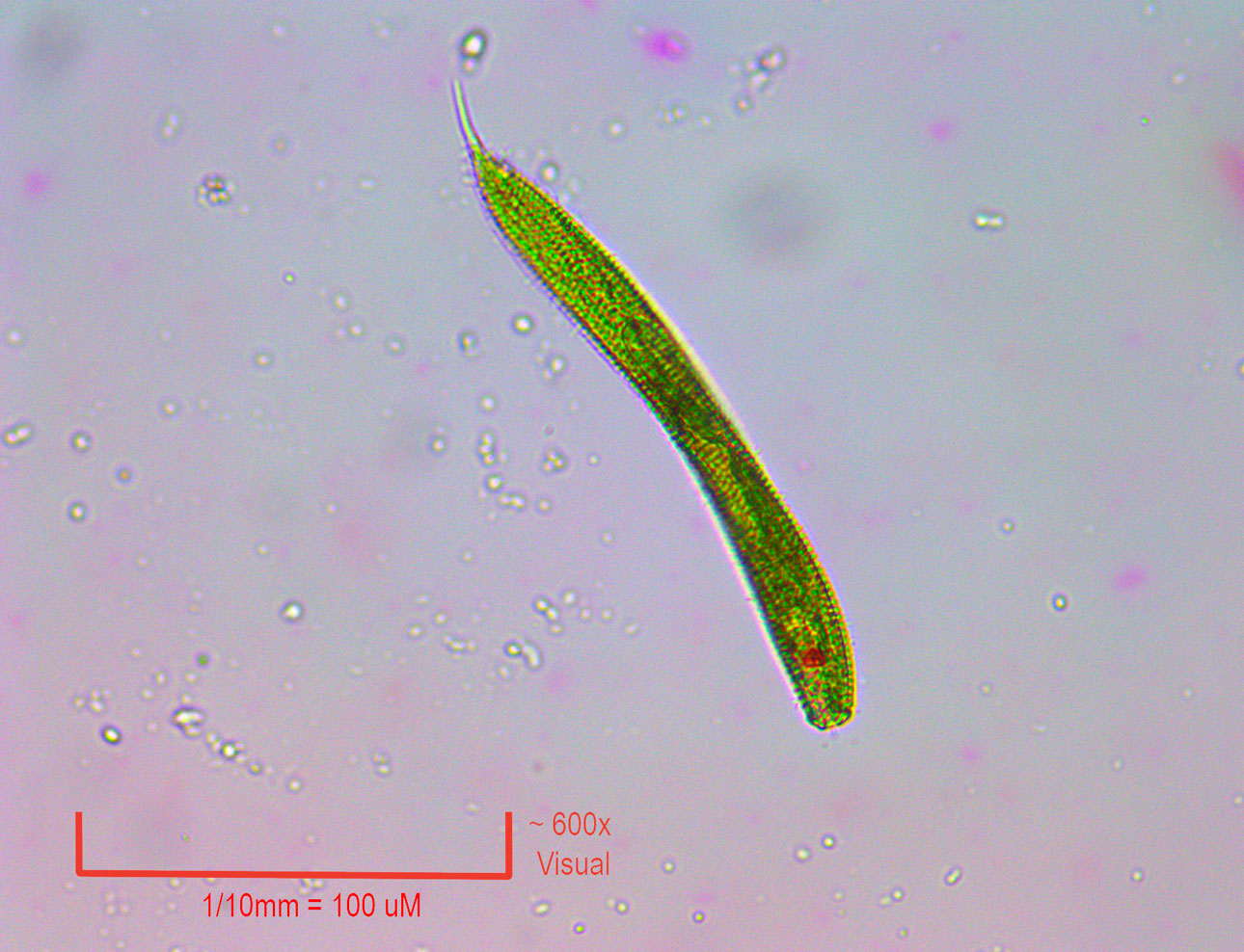
A
Euglenoid protozoan. at 150x. We have never found one of these
before! The green color is from organells which perform photosynthesis.
This is half way between plant and animal. It moved slowly, so
was easer to shoot. |
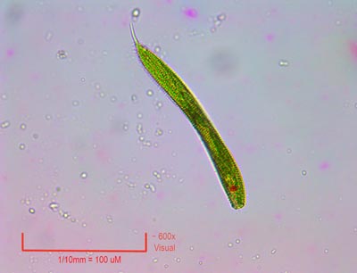 |
600x
on the Euglenoid. The red spot is its eye spot, as any organism
which can move and requires the light for food has a distinct
advantage if it can swim toward the light. |
 |
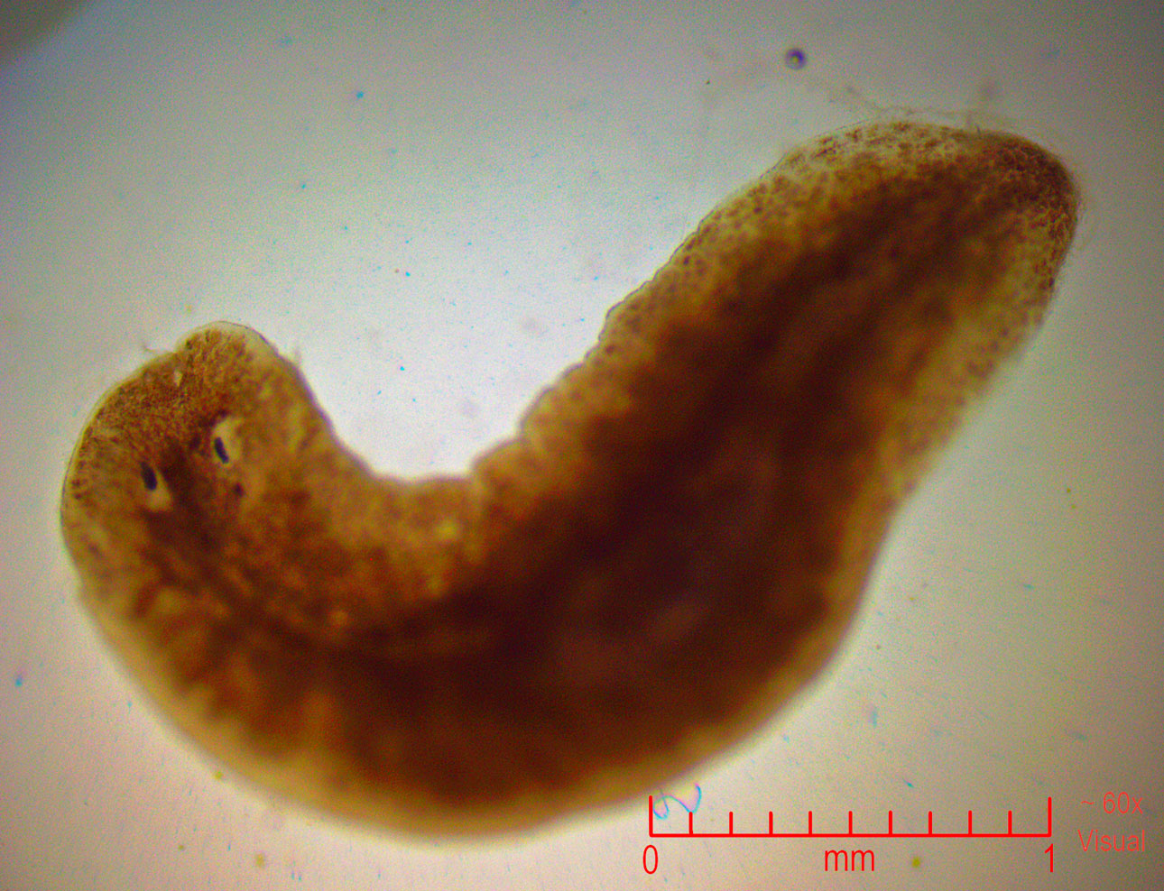
I
found this fellow on the bottom of one of the Duck weed petals
60x. He (It?) was creeping along looking for something to eat.
This is a Planarian flat worm. The first one we have ever found.
Note the eye spots inside the sunken cavities. While the worm
cannot actually form images, it can accurately sense the direction
of the light. |
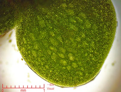 |
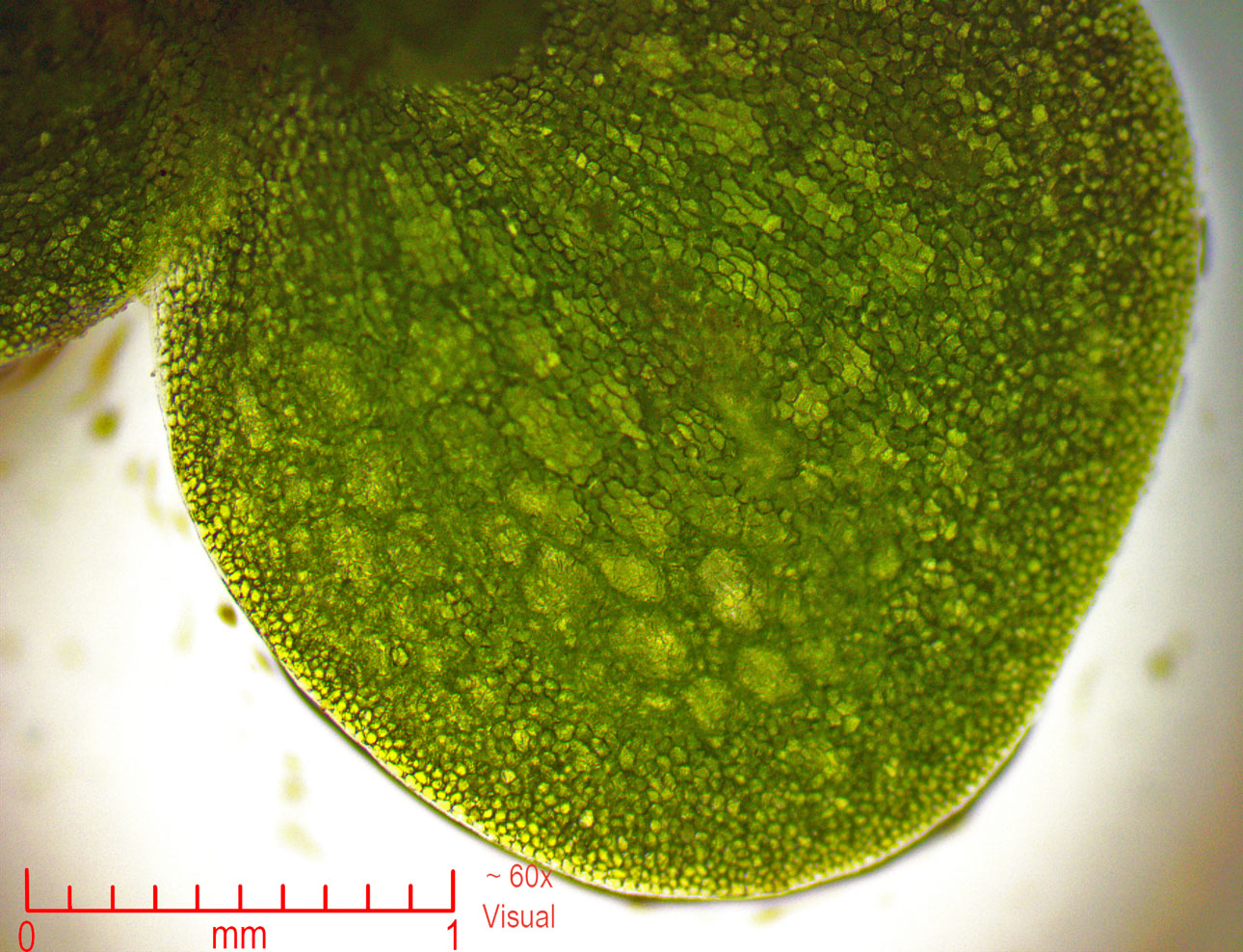
One
petal of a four leaf Duck weed at 60x. Individual cells on the
BOTTOM side here. This is the side that is in the water on this
floating mini plant. The other side has the stomata for air exchange. |
 |
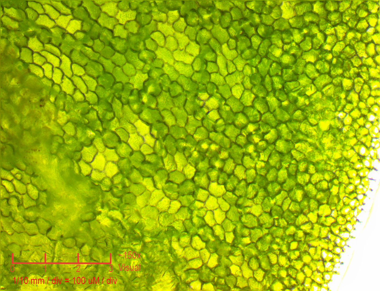
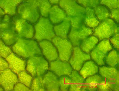
Closer
at 150x we see the jelly like cells more clearly. |
 |
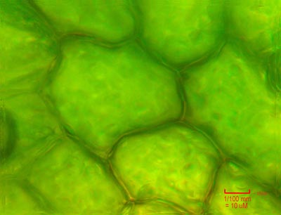
At
600x, the cells look like little nested pillows. |
 |
Finally,
at 1500x with the oil immersion lens, we can see the finest details
in the jelly like cells. |
|
Camera:  10 Megapixel CMOS
Platform:  AmScope Trinocular 2000x
Filters:  NONE
Location:  Payson, Az
Elevation:  5150 ft.
Processing: Photoshop  CS Pro
HOME
|